The New Standard of Care in Implant Dentistry
In the rapidly evolving landscape of modern dentistry, the transition from analog to digital is no longer a luxury—it is a clinical necessity. For the premium dental laboratory and the forward-thinking clinician, the integration of 3D-printed surgical guides represents the pinnacle of this evolution. As we move toward a future defined by predictability, the focus has shifted from simply 'using' digital tools to mastering the nuances of surgical guide accuracy and workflow integration.
Recent data suggests that the standard of care now requires clinicians to translate a digitally planned trajectory into the oral cavity with a level of precision that manual placement simply cannot replicate. With 3D-printed guides, we are seeing coronal deviations kept below 0.5 mm and angular errors maintained under 3°. This level of accuracy is not just about clinical success; it is about meeting the legal and professional expectations of 'reasonable accuracy' in a litigious medical environment.
The Anatomy of Accuracy: SLA vs. DLP Technologies
The foundation of a precise surgical guide lies in the technology used to fabricate it. While both Stereolithography (SLA) and Digital Light Processing (DLP) are prevalent in the industry, understanding their differences is crucial for dental technicians aiming for sub-millimeter precision.
- SLA (Stereolithography): Utilizes a laser to trace and cure resin. While known for smooth surface finishes, the point-by-point curing process can sometimes lead to longer print times.
- DLP (Digital Light Processing): Uses a projector screen to cure an entire layer of resin at once. This often results in faster production speeds and, in many modern units, exceptional dimensional stability for multi-unit guides.
Research indicates that additive-manufactured guides, particularly those utilizing sleeve-free designs, achieve the lowest crestal and apical deviations. By eliminating the mechanical tolerance required for metal sleeves, clinicians can achieve better angular control, ensuring the implant is placed exactly where the restorative plan dictates.
The Integrated Digital Workflow: From Scan to Surgery
A surgical guide is only as accurate as the data that informs its design. A truly integrated digital workflow relies on the seamless merging of two primary data sets: the surface anatomy and the underlying bone structure.
1. Data Acquisition and Merging
The process begins with high-resolution intraoral scanning (IOS) to capture the dentition and soft tissue, paired with Cone Beam Computed Tomography (CBCT) to visualize the bone and vital structures. The integration of these files (typically STL and DICOM) allows for a 'virtual patient' model. Any error at this stage—such as patient movement during the CBCT or a distorted IOS—will propagate through the entire workflow.
2. Restorative-Driven Planning
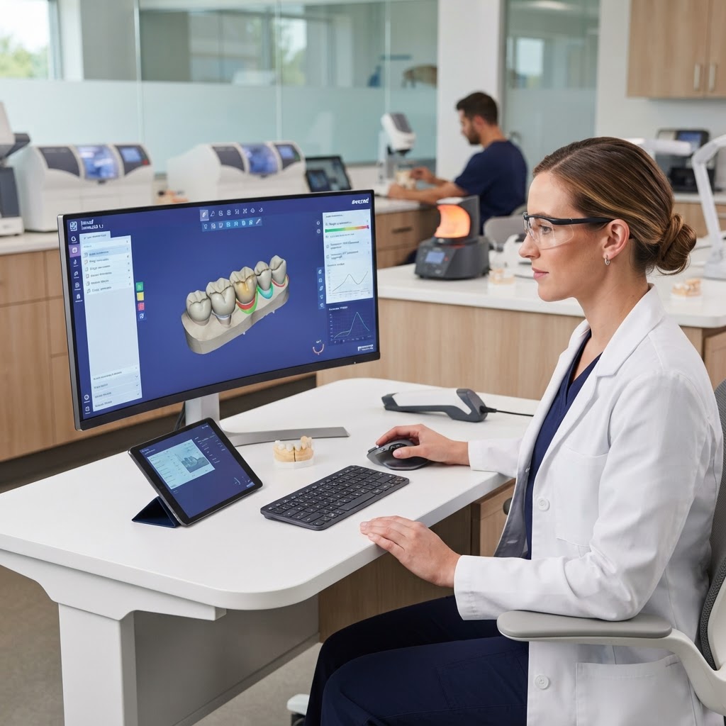
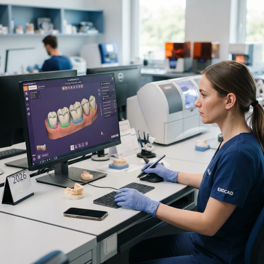
Modern CAD/CAM software allows us to plan the implant position based on the final prosthetic outcome. By visualizing the virtual crown first, we ensure that the implant is not only biologically integrated but also restoratively functional. This 'top-down' approach is the hallmark of a premium digital laboratory.
3. 3D Printing and Post-Processing
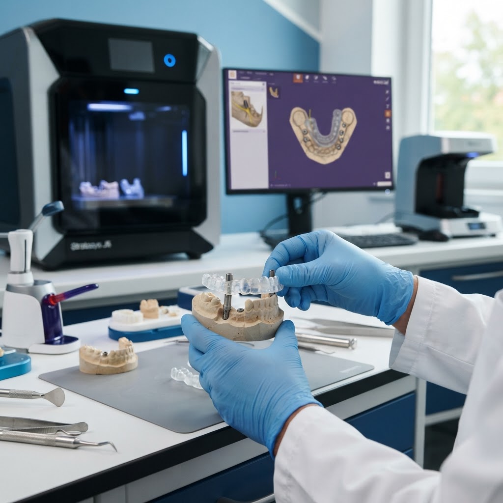
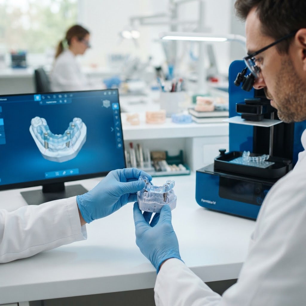
Once the design is finalized, the guide is printed using biocompatible resins. However, the workflow does not end at the printer. Proper post-processing—including thorough washing in isopropyl alcohol and calibrated UV curing—is essential to ensure the material reaches its full mechanical properties and maintains its dimensional accuracy. Failure to follow manufacturer protocols during curing can lead to guide warping, which compromises the fit on the patient's dentition.
Practical Insights for Labs and Clinics
For practices looking to integrate these technologies, the economic and clinical benefits are clear. In the United States, a custom 3D-printed surgical guide typically ranges from $150 for single-tooth cases to over $600 for complex full-arch rehabilitations. While the initial investment in hardware and software is significant, the ROI is found in reduced chair time and the elimination of costly surgical complications.
- Sleeve-Free Innovation: Consider adopting sleeve-free guide designs to minimize positional errors and reduce the bulk of the guide, improving visibility during surgery.
- AI-Driven Planning: Leverage emerging AI tools within planning software to automate nerve mapping and initial implant positioning, which can significantly reduce design time for the lab.
- In-House vs. Outsourced: While in-house printing offers faster turnaround, partnering with a premium digital lab like Cosmetic Smile Studio ensures access to high-end industrial printers and expert design oversight that may be difficult to maintain in a busy clinical setting.
The Future: AI and Dynamic Navigation
The next frontier in implant dentistry involves the marriage of static 3D-printed guides with dynamic navigation and AI. Artificial intelligence is already being used to enhance the merging of CBCT and IOS data, reducing the risk of human error in the planning phase. Furthermore, the integration of augmented reality (AR) may soon allow clinicians to visualize the planned implant position through the surgical guide in real-time, providing an extra layer of safety and precision.
Conclusion
Surgical guide accuracy is the bridge between a brilliant digital plan and a successful clinical reality. By mastering the digital workflow—from precise data acquisition to calibrated 3D printing—dental professionals can provide a level of care that is safer, faster, and more predictable than ever before. As the industry moves toward a fully digital ecosystem, staying informed on the latest material sciences and integration strategies is paramount.
Ready to elevate your implant precision? Contact Cosmetic Smile Studio today to learn how our advanced digital workflow and premium surgical guide fabrication can transform your practice's surgical outcomes.