The Evolution of Precision: The Digital Mandate in Modern Implantology
In the contemporary dental landscape, the transition from freehand implant placement to fully guided surgery represents one of the most significant shifts in clinical protocol. No longer viewed as a luxury for complex cases, surgical guides have become the cornerstone of predictable, restoratively driven outcomes. For the premium digital laboratory and the high-end surgical clinic, the focus has shifted from the mere adoption of the technology to the relentless pursuit of micron-level accuracy. Achieving this requires a deep understanding of the digital thread—from the initial scan to the final light-cure. When we discuss surgical guide printing accuracy, we are not just discussing hardware; we are discussing a holistic integration of data, material science, and mechanical engineering.
The Foundation: High-Fidelity Data Acquisition and Alignment
The accuracy of a surgical guide is fundamentally limited by the quality of the input data. The digital workflow begins with the marriage of hard and soft tissue data, typically through the fusion of DICOM files from Cone Beam Computed Tomography (CBCT) and STL/PLY files from Intraoral Scans (IOS). Any discrepancy during this alignment phase propagates through the entire workflow, leading to a guide that may seat perfectly but directs the osteotomy into an unintended trajectory.
The Role of Radiographic Templates
In edentulous cases, the use of radiographic templates with fiducial markers remains the gold standard. This allows for a precise 'dual scan' protocol that eliminates the common artifacts associated with large metal restorations. For partially edentulous patients, the laboratory must ensure that the intraoral scan captures sufficient tooth morphology and soft tissue detail to allow for a stable, 'snap-fit' guide. Even a 0.1mm deviation in the virtual alignment can result in several degrees of angular deviation at the apex of a long implant.
Printer Selection: Navigating the Hardware Landscape
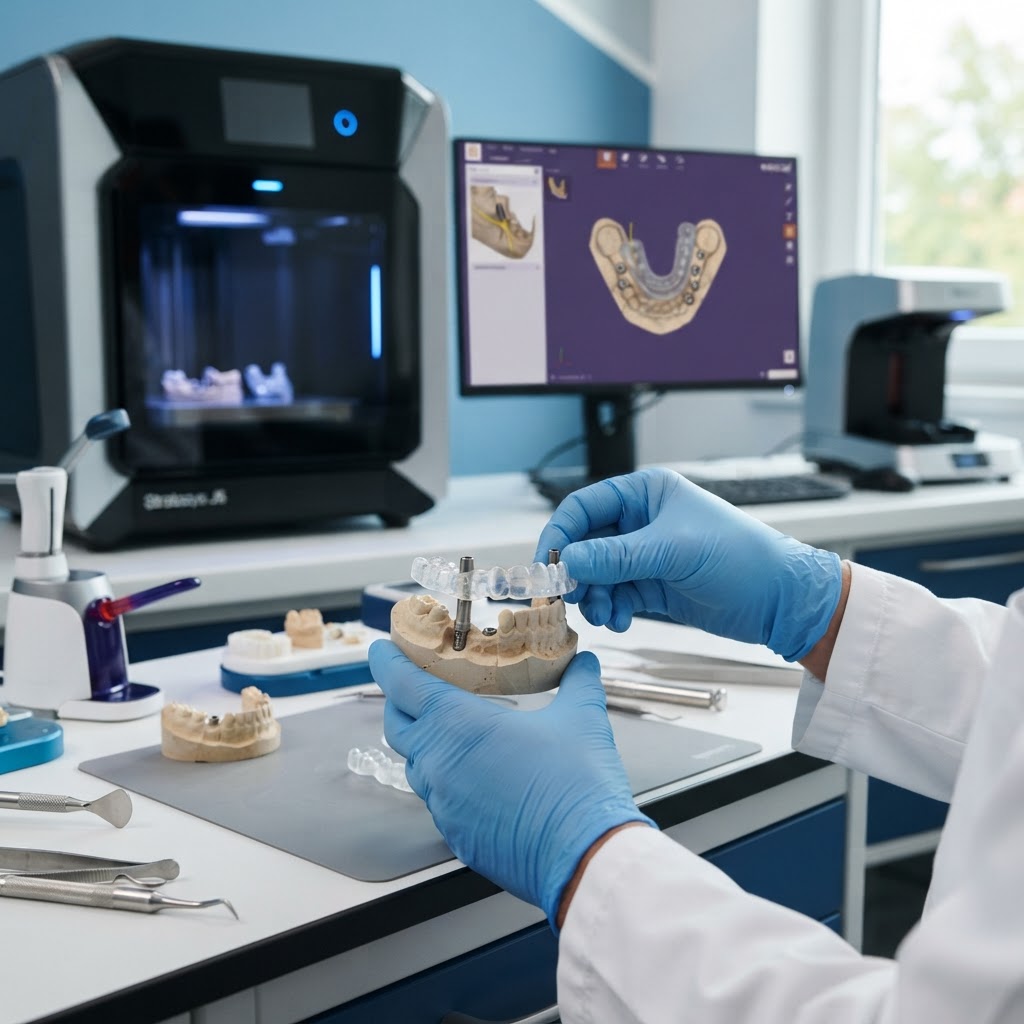
When it comes to the fabrication of surgical guides, the choice of 3D printing technology is paramount. While various technologies exist, the industry has largely converged on three main modalities: Digital Light Processing (DLP), Stereolithography (SLA), and Masked Stereolithography (mSLA/LCD).
- DLP (Digital Light Processing): Known for its speed and exceptional accuracy, DLP uses a projector to cure an entire layer of resin at once. Because it uses a pixel-based projection, laboratories must be mindful of 'pixelation' effects, though modern high-resolution projectors have largely mitigated this.
- SLA (Stereolithography): This uses a laser to draw each layer. While historically slower than DLP, SLA provides incredibly smooth surface finishes and high dimensional stability, which is critical for ensuring the surgical sleeve fits perfectly without binding.
- LCD/mSLA: The most accessible of the three, LCD printers use an LED array and an LCD mask. Recent advancements in monochrome LCD screens have brought the accuracy and durability of these machines into the professional sphere, making them a viable option for high-volume guide production.
The Criticality of Design Parameters and Sleeve Calibration
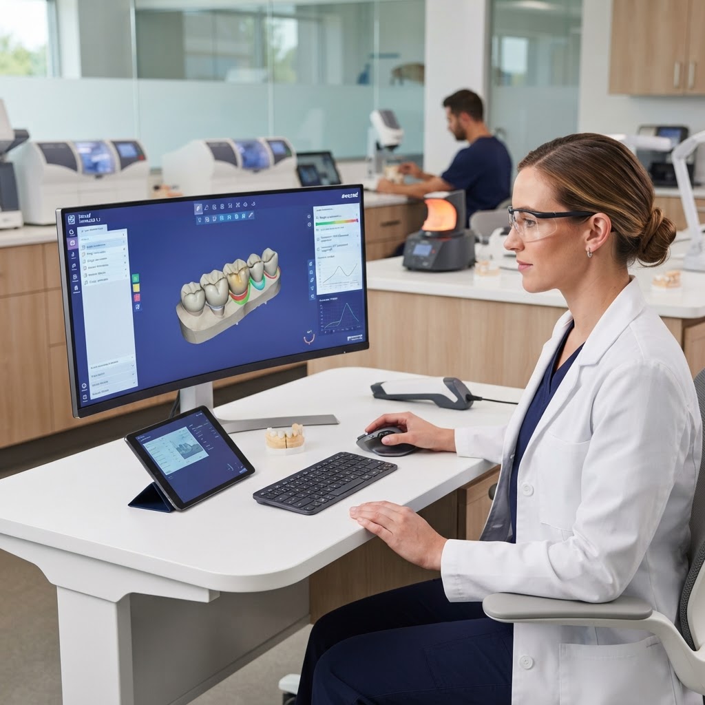
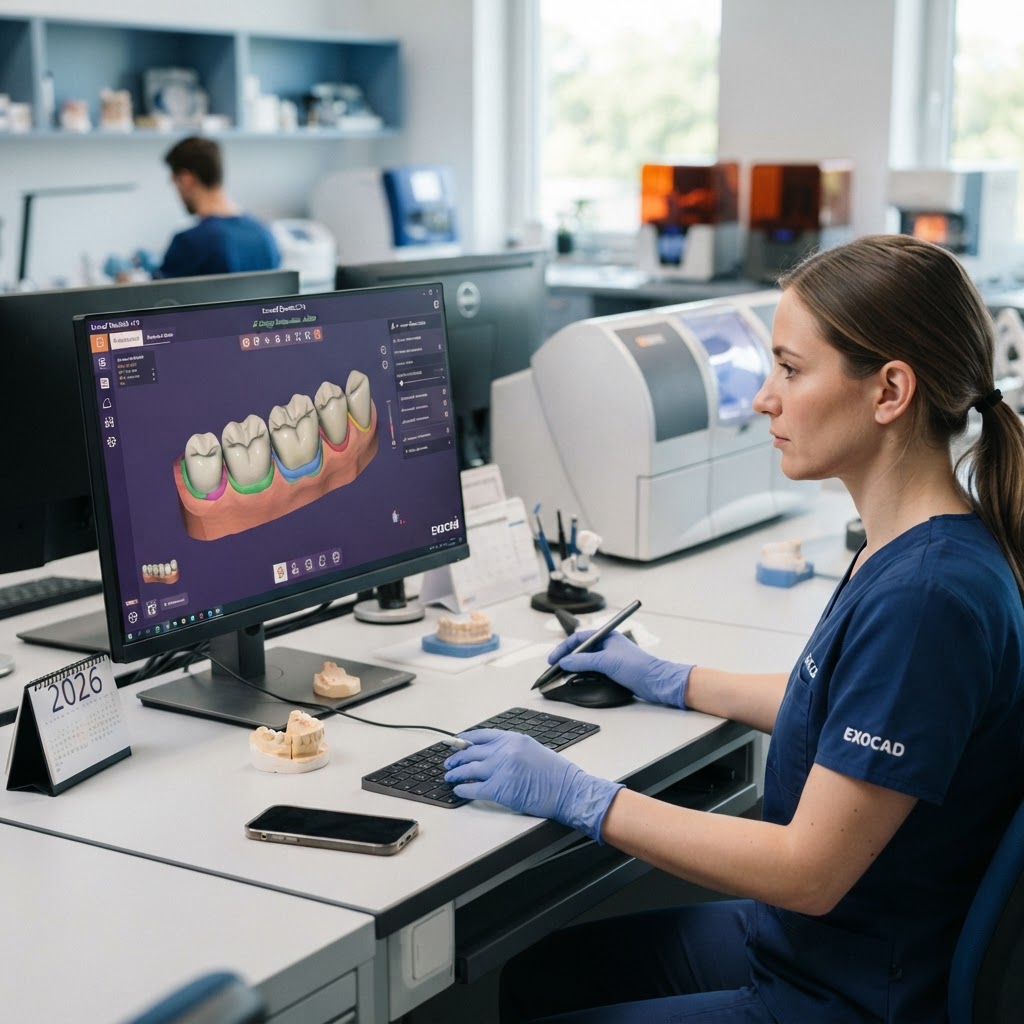
The design phase in software like exocad or 3Shape is where the 'fit' is determined. A common mistake is using a one-size-fits-all approach to guide offsets. The 'offset'—the space between the printed guide and the surgical sleeve—must be calibrated to the specific printer and resin combination being used.
- Optimal Wall Thickness: For surgical guides, a wall thickness of 2.5mm to 3.0mm is generally recommended to prevent flexing during the surgical procedure.
- Offset Calibration: A standard starting point is often 0.05mm to 0.1mm, but this must be validated through test prints. If the sleeve is too tight, it may crack the guide during insertion; if too loose, the drill's trajectory is compromised.
- Inspection Windows: Integrating large windows into the guide design allows the clinician to visually confirm that the guide is fully seated on the occlusal surfaces of the teeth.
Material Science: Biocompatibility and Dimensional Stability
Surgical guides require specialized Class I or Class VI biocompatible resins. These materials are engineered to withstand the friction and heat generated during the drilling process without off-gassing or degrading. However, these resins are also prone to shrinkage and warping if not handled correctly. Modern dental resins are formulated with photo-initiators that react to specific wavelengths (usually 385nm or 405nm). It is critical that the laboratory uses the 'validated' settings provided by the resin manufacturer for their specific printer. Deviating from these settings, even by a few seconds of exposure time, can result in a guide that is either brittle or dimensionally inaccurate.
Post-Processing: The Final Micron
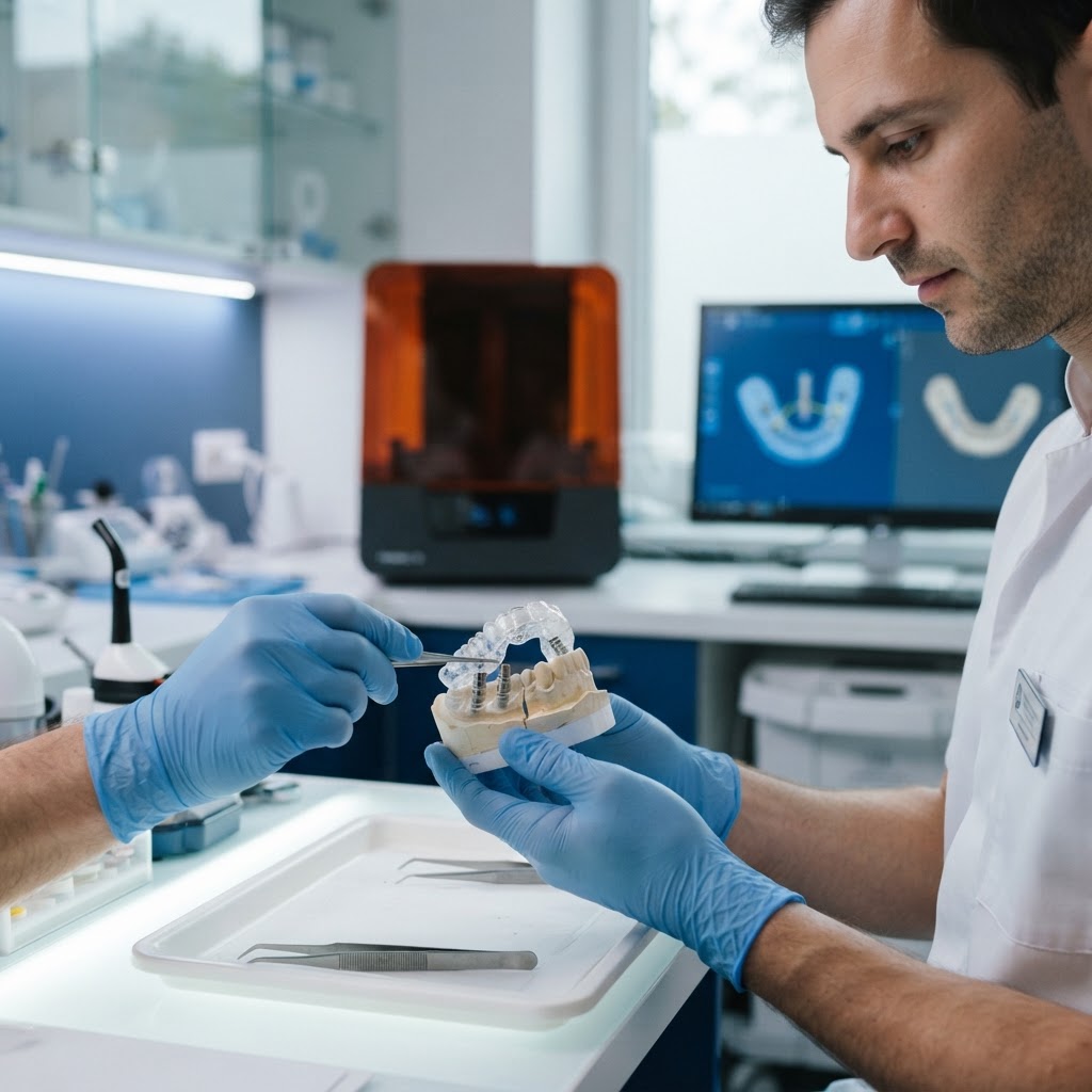
Perhaps the most overlooked aspect of surgical guide accuracy is the post-processing stage. The accuracy of a print can be completely undone by improper washing or curing.
- Validated Washing: Over-soaking a guide in Isopropyl Alcohol (IPA) can lead to material softening and swelling, which permanently alters the internal geometry. Use an automated wash system and follow the manufacturer's timing precisely.
- The Curing Cycle: Proper polymerization is essential for both biocompatibility and strength. Using a high-end, nitrogen-purged curing unit or a temperature-controlled UV box ensures that the resin reaches its maximum cross-link density, preventing 'creep' or warping before the surgery.
- Sleeve Insertion: Sleeves should be inserted using a calibrated press. If the design and print are accurate, the sleeve should seat with a firm, tactile 'click.'
Conclusion: The Path to Clinical Excellence
The successful integration of surgical guide printing into a digital workflow is not merely about purchasing a 3D printer; it is about mastering the variables that influence the final product. By focusing on high-fidelity data acquisition, calibrated hardware settings, and rigorous post-processing protocols, dental laboratories can provide clinicians with the confidence to perform complex surgeries with unprecedented precision. As we move further into 2026, the gap between traditional and guided surgery will continue to widen, with the latter becoming the undeniable standard for patient safety and restorative success. For labs ready to lead this charge, the investment in accuracy is an investment in the future of the profession. Contact Cosmetic Smile Studio today to learn how our advanced digital workflows can elevate your implant cases.