The Evolution of Precision in Modern Implantology
In the contemporary dental landscape, the transition from freehand implant placement to guided surgery is no longer a luxury—it is a standard of care. As clinicians and dental laboratories strive for the 'perfect' placement, the focus has shifted from the mere feasibility of the procedure to the micron-level accuracy of the tools involved. Surgical guide printing accuracy is the linchpin of this evolution, bridging the gap between virtual treatment planning and clinical reality.
For the modern dental professional, understanding the nuances of digital workflow integration is essential. It is not enough to simply own a 3D printer; one must master the synergy between data acquisition, software manipulation, and material science to ensure that the final guide provides the predictable results patients expect.
The Pillars of Surgical Guide Accuracy
Accuracy in guided surgery is cumulative. Errors introduced at any stage of the digital workflow—from the initial scan to the final light-cure—can compound, leading to significant deviations at the implant apex. To achieve high-fidelity results, we must address three primary pillars:
- Data Fidelity: The foundation of any surgical guide is the alignment of DICOM data from CBCT imaging and STL data from intraoral scanners. Any distortion in the soft tissue representation or radiographic artifacts can compromise the fit.
- Software Calibration: CAD software must account for the specific 'offset' or 'tolerance' required for the surgical sleeves. This varies depending on the implant system and the specific 3D printer used.
- Dimensional Stability: The choice of resin and the printing technology (SLA, DLP, or LCD) directly impact the guide's resistance to deformation during the surgical procedure and sterilization.
Optimizing the Digital Workflow Integration
A seamless digital workflow is characterized by the frictionless transfer of data between the clinic and the laboratory. At Cosmetic Smile Studio, we emphasize a 'closed-loop' approach to minimize manual intervention and human error.
Phase 1: Advanced Data Acquisition
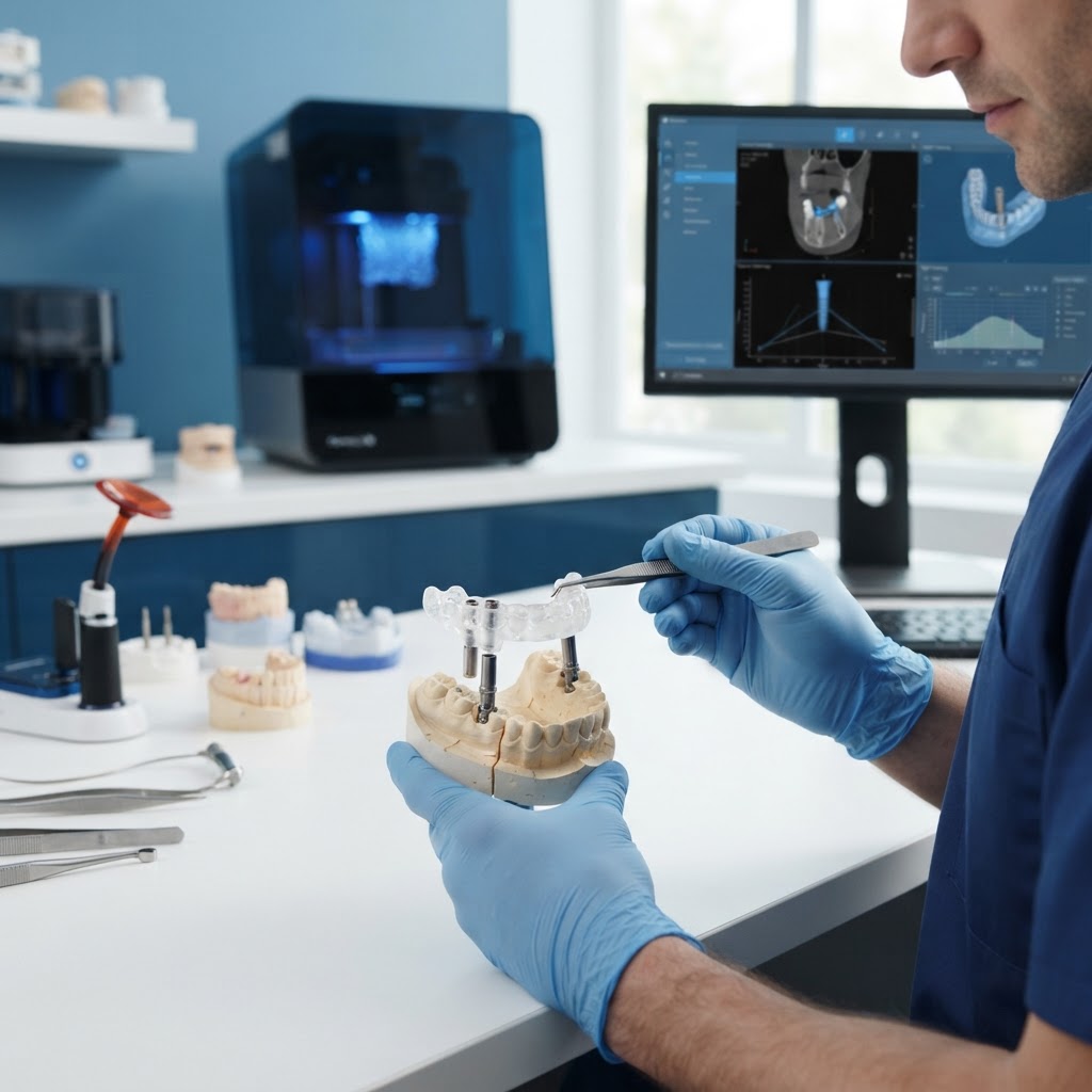
Success begins with a high-resolution intraoral scan. Clinicians should ensure that the scan captures sufficient tooth morphology and stable soft tissue landmarks. When integrating these with CBCT data, using a radiographic template for edentulous cases can significantly enhance the precision of the coordinate system alignment.
Phase 2: Virtual Planning and Sleeve Selection
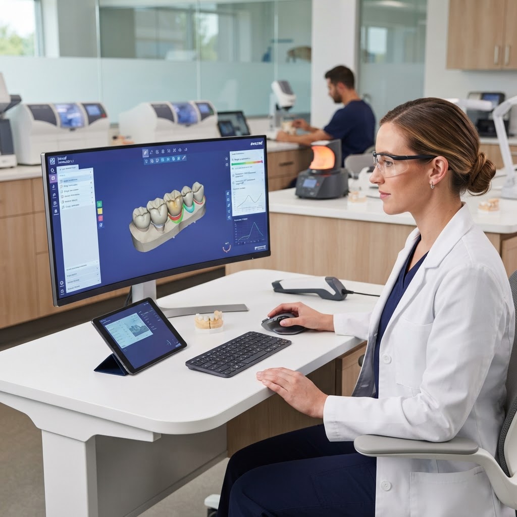
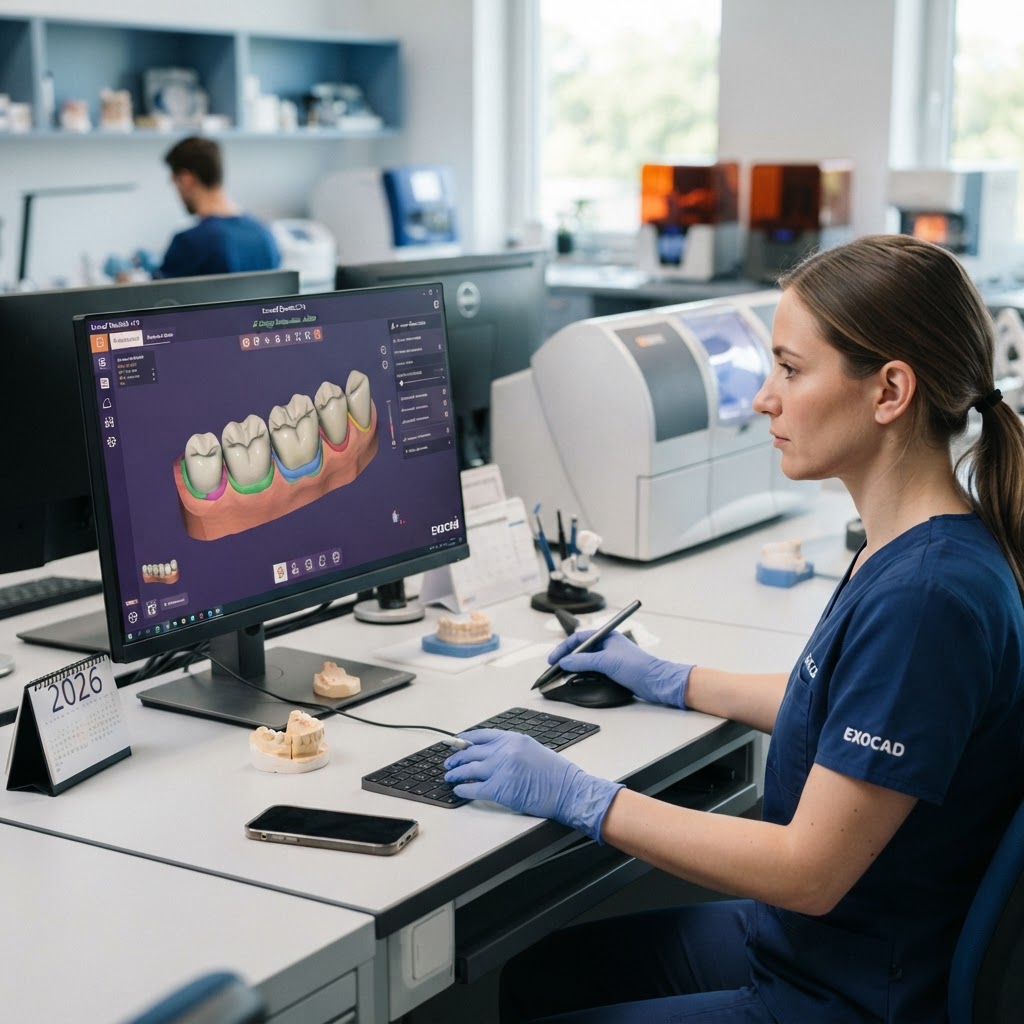
The transition from a prosthetic-driven plan to a surgical reality requires precise sleeve positioning. It is critical to select the correct offset in the planning software (such as exocad or 3Shape). Even a 0.1mm discrepancy in the software's internal diameter settings can lead to a guide that is either too tight to seat the drill or too loose to maintain trajectory.
Phase 3: High-Resolution 3D Printing
While various 3D printing technologies exist, Digital Light Processing (DLP) and Liquid Crystal Display (LCD) have become the industry standards for surgical guides due to their speed and XY resolution. However, the accuracy of a print is heavily dependent on the layer height and the orientation of the guide on the build plate. Printing at a 45-degree angle often provides the best balance between surface finish and dimensional accuracy, reducing the 'stair-stepping' effect on critical fit surfaces.
The Impact of Material Science and Post-Processing
The biocompatible resins used for surgical guides are engineered for rigidity and clarity. However, these materials are sensitive to environmental factors. Over-curing can lead to brittleness and shrinkage, while under-curing leaves residual monomers that compromise biocompatibility and strength.
- Wash Protocols: Using high-purity Isopropyl Alcohol (IPA) for the correct duration is vital. Excessive soaking can soften the resin and cause the guide to warp.
- Automated Curing: Using a validated UV curing chamber ensures that the guide reaches its full mechanical properties. This step is non-negotiable for maintaining the tight tolerances required for surgical sleeves.
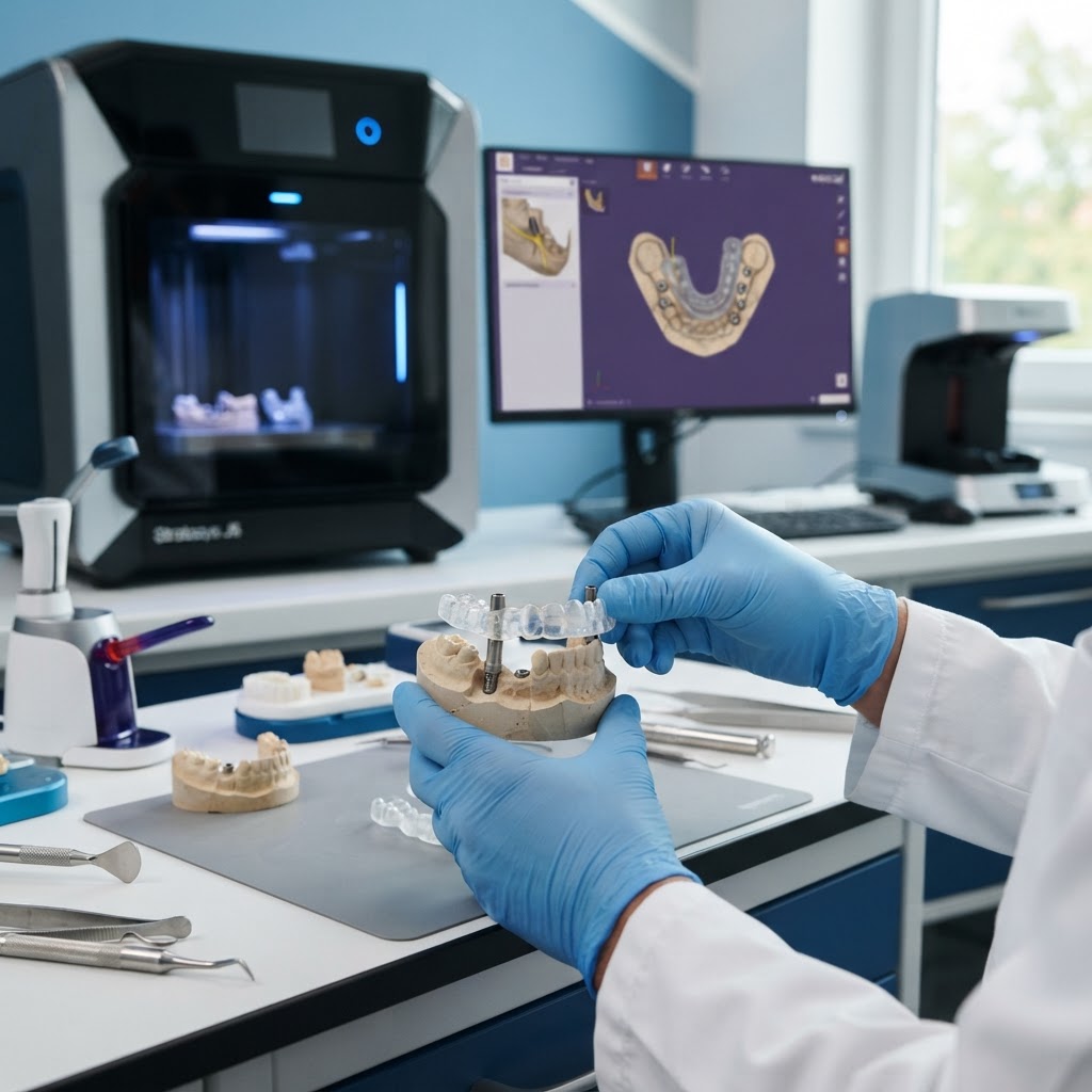
- Fit Verification: Every guide should be verified on a 3D-printed model of the patient’s arch before the surgery. If the guide rocks or shows gaps, the workflow must be re-evaluated for discrepancies.
Common Pitfalls and Professional Insights
In our experience at the laboratory, the most common cause of guide failure is not the printer itself, but the lack of 'fit compensation' in the design phase. As resins shrink slightly during the curing process, the CAD design must account for this predictable change. Furthermore, the heat generated during the drilling process can cause thermal expansion in lower-quality resins; therefore, choosing premium, heat-resistant biocompatible materials is essential for long, complex surgeries.
The Future of Guided Workflows: AI and Real-Time Tracking
Looking ahead, we are seeing the integration of Artificial Intelligence to automate the segmentation of CBCT data, further reducing the margin for error. Additionally, the rise of dynamic navigation—where the digital plan is tracked in real-time by cameras—complements 3D-printed guides by providing a secondary layer of verification during the osteotomy.
Conclusion: Partnering for Precision
Achieving surgical guide printing accuracy is a meticulous process that rewards attention to detail. By integrating high-resolution data, calibrated software, and validated post-processing, dental practices can virtually eliminate the risks associated with implant placement. At Cosmetic Smile Studio, we specialize in bridging the gap between digital design and clinical excellence.
Ready to elevate your implant outcomes? Contact our technical team today to discuss how our high-precision surgical guide services can integrate into your existing digital workflow. Let us help you provide the predictable, life-changing results your patients deserve.